Colon Scaffold
The current colon scaffold is 3D Colon 1 built from class MeshType_3d_colon1.
The human variant is shown in Fig. 76.
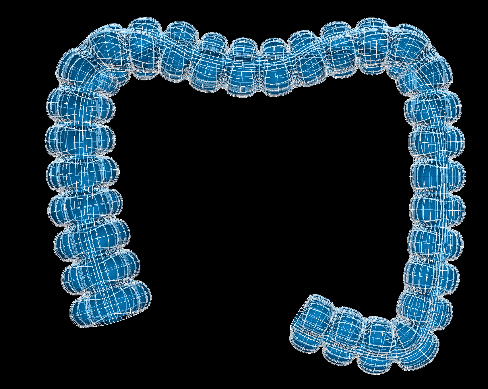
Fig. 76 Human colon scaffold.
The colon scaffold is a 3-D volumetric model of the colon representing the proximal, transverse and distal colon.
Variants
The colon scaffold is provided with parameter sets for the following four species, which differ in shape, and in particular have different numbers of tenia coli:
Cattle (no tenia coli)
Human (3 tenia coli)
Mouse (no tenia coli)
Pig (2 tenia coli)
These variants’ geometry and annotations are best viewed in the Scaffold Creator tool in the ABI Mapping Tools.
On the web, the latest published generic colon scaffold variants can be viewed on the
SPARC Portal by searching for colon, filtering for anatomical models, selecting a
variant and viewing the scaffold in its Gallery tab or via the Organ Scaffolds help article.
The colon scaffold script generates the scaffold mesh and geometry from an idealization of their cross-sectional profile shapes. The mesh is derived from ellipsoid and cubic functions based on a one dimensional central path which describes the path of the colon. The parameters were carefully tuned for each species, and it is not recommended that these be edited.
Instructions for editing the central path are given with the ABI Mapping Tools Scaffold Creator documentation. Note that the D2 derivative along the path points towards the first node around the cross-section along the colon. If editing, use the Interactive Functions to Smooth derivatives, and Smooth side cross derivatives to make these as smooth as required.
The central path used to generate the current mouse colon scaffold is obtained from tracing a central path from a dissection image obtained from the literature. Lixin Wang (UCLA), Yvette Tache (UCLA), and Marthe Howard (University of Toledo) provided lengths and diameter data for mouse colon. The pig colon scaffold is generated from images and measurements provided by Million Mulugeta, Muriel Larauche, and Yvette Tache (UCLA). The human colon scaffold is created based on average dimensions and images obtained from the literature.
The mucosa, submucosa, circular muscle, longitudinal muscle and serosa layers of the colon are fully represented on
the scaffold when Number of elements through wall is set to 4. Alternatively, the entire colon wall can be
represented as a single layer by setting Number of elements through wall to 1.
Coordinates
The colon scaffold defines the geometric, flat and material coordinates.
The geometric coordinates field gives an approximate, idealized representation of the colon shape for the species,
which is intended to be fitted to actual data for a specimen.
The flat coordinates represents the geometric field when the colon scaffold is cut along its length and laid flat.
This field is intended for fitting data obtained from a flat colon preparation.
The material coordinates field colon coordinates defines a highly idealized coordinate system to give permanent
locations for embedding structures in the colon. It is a cylindrical tube and can be viewed by visualising this field in
the Display tab of Scaffold Creator.
The colon scaffold supports limited refinement/resampling by checking Refine (set parameter to true) with chosen
Refine number of elements parameters. Be aware that only the coordinates field is currently defined on the refined
mesh (but annotations are transferred).
Annotations
Important anatomical regions of the colon are defined by groups of elements (or faces, edges and nodes/points) and annotated with standard term names and identifiers from a controlled vocabulary.
Annotated 3-dimensional volume regions are defined by groups of 3-D elements including:
ascending colon (only in human)
circular muscle layer of colon
colon
colonic mucosa
descending colon (only in human)
distal colon (in cattle, mouse, pig)
longitudinal muscle layer of colon
proximal colon (in cattle, mouse)
spiral colon (only in pig)
submucosa of colon
tenia coli (in human, pig)
tenia libera (only in human)
tenia mesocolica (only in human)
tenia omentalis (only in human)
transverse colon
Terms for volume regions such as the above are not to be used for digitized contours! They are used for applying different material properties in models and the strain/curvature penalty (stiffness) parameters in fitting.
Annotated 2-dimensional surface regions are defined for matching annotated contours digitized from medical images
including (where surface is the outside boundary on the meshes):
luminal surface of the colonic mucosa
serosa of colon