Brainstem Scaffold
The current brainstem scaffold is 3D brainstem 1 built from class MeshType_3d_brainstem1.
The human variant is shown in Fig. 75.
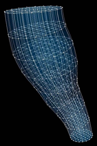
Fig. 75 Human brainstem scaffold.
The brainstem scaffold is a 3-D volumetric model of the brainstem representing all three parts: midbrain, pons, and medulla oblongata. Each part is meshed with its own elements but these are connected with common faces to the elements of the neighbouring parts.
Variants
The brainstem scaffold is provided with parameter sets for the following six species, which differ in shape and size.
Cat
Human
Mouse
Pig
Rat
Sheep
These variants’ geometry and annotations are best viewed in the Scaffold Creator tool in the ABI Mapping Tools.
On the web, the latest published generic brainstem scaffold variants can be viewed on the
SPARC Portal by searching for brainstem, filtering for anatomical models, selecting a
variant and viewing the scaffold in its Gallery tab or via the Organ Scaffolds help article.
The brainstem scaffold script generates the scaffold mesh and geometry from a solid cylinder function based on a one dimensional central path with side axes controlling lateral dimensions. The parameters were carefully tuned for each species, and it is not recommended that these be edited.
Instructions for editing the central path are given with the ABI Mapping Tools Scaffold Creator documentation. Note that the D2 and D3 derivatives control the side dimensions, and derivatives D12 and D13 control the rate of change of these along the central path. If editing, use the Interactive Functions to Smooth derivatives, Make side derivatives normal and Smooth side cross derivatives to make these as smooth as required.
The generic brainstem scaffolds are fitted and smoothed to the segmentation data from a human (BodyParts3D V4.3i), a pig (Beckman Institute for Advanced Science and Technology, Pig Imaging Group, University Of Illinois Urbana-Champaign), a rat (NeuroRat V4.0), and a sheep (Nitzsche et al.).
Note
The central path is annotated with midbrain, pons, and medulla oblongata regions but these are only
approximately used to define their geometric extents in the final scaffold. Be aware that material coordinates are
only correctly defined when there are exactly 8 elements along the brainstem. These limitations will be fixed at a
later date.
Coordinates
The brainstem scaffold defines both geometric and material coordinates.
The geometric coordinates field gives an approximate, idealized representation of the brainstem shape for the
species, which is intended to be fitted to actual data for a specimen.
The material coordinates field brainstem coordinates defines a highly idealized coordinate system to give
permanent locations for embedding structures in the brainstem. This is a cylinder of unit radius and 8 units long:
intended to be 3 units for medulla oblongata, 3 units for pons, and 2 units for midbrain. Be aware at this time that
these ratios are only correctly defined if there are 8 elements along the brainstem.
The brainstem scaffold supports limited refinement/resampling by checking Refine (set parameter to true) with
chosen Refine number of elements~ parameters. Be aware that only the coordinates field is currently defined
on the refined mesh (but annotations are transferred).
Annotations
Important anatomical regions of the brainstem are defined by groups of elements (or faces, edges and nodes/points) and annotated with standard term names and identifiers from a controlled vocabulary.
Annotated 3-dimensional volume regions are defined by groups of 3-D elements:
brainstem
medulla oblongata
midbrain
pons
Terms for volume regions such as the above are not to be used for digitized contours! They are used for applying different material properties in models and the strain/curvature penalty (stiffness) parameters in fitting.
Annotated 2-dimensional surface regions are defined for matching annotated contours digitized from medical images
including (where interface means the outside boundary of the brainstem connecting to different organs such as
thalamus and spinal cord, exterior is the outside boundary of the brainstem not including interfaces to structures
along its length):
brainstem exterior
brainstem-spinal cord interface
medulla oblongata exterior
midbrain exterior
pons exterior
thalamus-brainstem interface
Annotated 1-dimensional line regions are defined on the central path and used to label elements in each part (but be aware of their limitations noted above):
medulla oblongata
midbrain
pons
Several fiducial marker points are defined on the brainstem scaffold, of which the following eight are potentially usable when digitizing:
brainstem dorsal midline caudal point
brainstem ventral midline caudal point
brainstem dorsal midline pons-medulla junction
brainstem ventral midline pons-medulla junction
brainstem dorsal midline midbrain-pons junction
brainstem ventral midline midbrain-pons junction
brainstem dorsal midline cranial point
brainstem ventral midline cranial point